Articolo del 10/07/2024
Magnetic resonance imaging is an advanced imaging technique that utilizes magnetic fields to create detailed images of organs and tissues within the body.
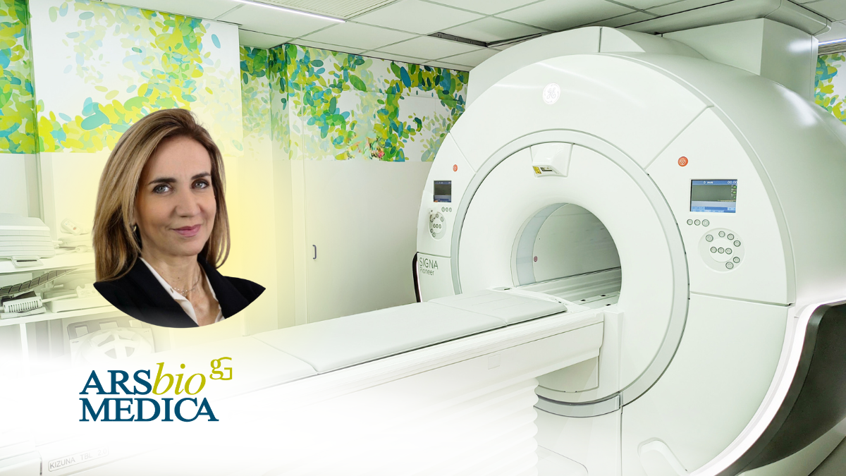
At Arsbiomedica, we perform high-field Magnetic Resonance Imaging (MRI) using 3 Tesla technology with Artificial Intelligence, which ensures significantly faster and more accurate diagnostic exams compared to previous technologies, offering a new standard of comfort for the patient.
Moreover, the use of a contrast agent, which is an injectable substance typically based on gadolinium, allows for a more precise study of certain structures during the MRI.
What are the advantages of performing MRI with 3 Tesla technology? When should MRI with contrast agent be performed?
Let’s explore the topic with Dr. De Cicco, a radiologist at Arsbiomedica.
Based on clinical indications, in some selected cases it is necessary to perform the MRI exam with the use of a contrast agent, which is administered intravenously: this involves the administration of a few cc (generally 8-10 cc) of Gd, a paramagnetic chemical element that enhances blood flow.
When is it necessary to perform MRI with contrast agent?
The contrast agent is crucial for clearly highlighting blood vessels, tumors, lesions, or inflammations that might otherwise not be visible in standard images.
- The administration of the contrast agent under the meticulous supervision of the Radiologist allows for a better evaluation of vascular structures in the brain, chest, abdomen, and limbs, enabling the identification of narrowings (stenoses), dilations (such as cerebral aneurysms), congenital vessel anomalies (vascular malformations known as AVMs) with better anatomical detail compared to a baseline assessment.
- The use of the contrast agent also allows for the study of perfusion distribution in organs, which can be essential, for example, in the brain in cases of ischemia, in the heart during a heart attack, or in various organs when infarct lesions are suspected, such as in the liver and spleen.
- Furthermore, the contrast agent is considered essential in the evaluation of a focal lesion, meaning when an incidental nodule is found in various organs through ultrasound or non-contrast MRI (in the brain, liver, pancreas, for example), it is crucial to determine its benign or malignant nature and to characterize the lesion as much as possible from an anatomical-pathological perspective.
How is the exam performed?
Before undergoing an MRI with contrast agent, after the Radiologist confirms the need for a contrast agent based on the clinical indication provided by the referring physician, the patient is assisted by an Anesthesiologist who conducts a targeted questionnaire to exclude any contraindications and to insert a peripheral vein cannula, usually in the arm.
The Anesthesiologist will continue to be present during the MRI exam until the contrast agent is administered.
Are there any contraindications?
The only contraindications are any previous allergic reactions to the same drug or other allergens, which must be thoroughly investigated, and renal insufficiency: renal function is particularly crucial when studying hepatic lesions and bile ducts because the contrast agent used is largely eliminated by the kidneys. Therefore, the patient will be asked to show recent creatinine levels (within the last 3 months).
How to prepare for the MRI?
The use of a contrast agent in MRI does not require any preparation: however, a light diet is advisable before the administration of the contrast agent, or better yet, fasting, simply because the patient must remain in a supine position, which can help avoid reflux and regurgitation of food during the exam.


